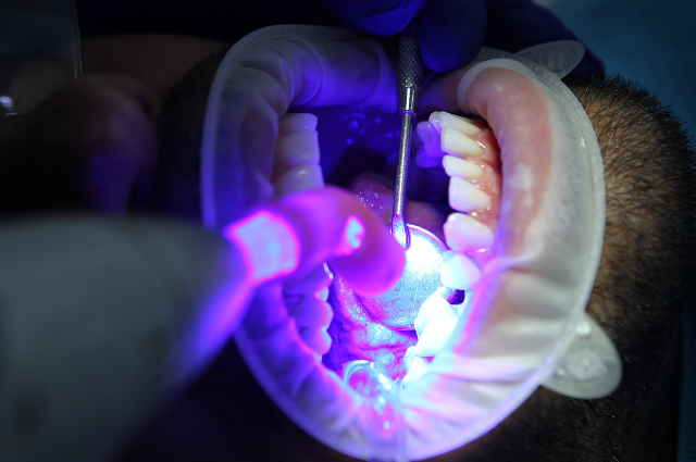
Image by diego toral from Pixabay
The field of stem cell research has greatly advanced our understanding of these cells as key contributors to organ formation and the maintenance of adult tissues since their concept emerged in the early 20th century. Their exceptional regenerative capabilities have also spurred the development of tissue engineering technologies, particularly during the late 20th century.
Stem cells are undifferentiated cells with the ability to self-renew through cell division and differentiate into specialized cells with specific functions. The term "stem cell" was introduced to describe cells that give rise to the germline.
In the context of dental tissues, several types of stem cell populations have been identified, including dental pulp stem cells, stem cells from human exfoliated deciduous teeth, apical papilla stem cells, dental follicle progenitor cells, and periodontal ligament stem cells. These cells are readily accessible, exhibit high plasticity and multipotential characteristics, and represent the gold standard for bone regeneration derived from neural crest cells. Additionally, they hold promise for safe, autologous therapeutic applications aimed at repairing tissue defects.
The accessibility of dental and periodontal tissues makes them appealing sources for isolating autologous mesenchymal stem cells (MSCs), offering a solution to the challenges posed by the invasive procedures required to harvest MSCs from other sources. Advances in isolating, collecting, and cryopreserving dental pulp progenitor cells for both storage and clinical use are now practical and commercially feasible.
Dental Stem Cells
Oral health is an important reflection of overall health, as highlighted by the World Health Organization (WHO). However, maintaining the integrity and vitality of dental tissues, especially the dental pulp, presents a significant challenge in modern dentistry. The dental pulp is essential for maintaining tooth health but is vulnerable to damage and infection, which can result in conditions such as irreversible pulpitis or necrosis.
Compared to other tissues like bone, human teeth have a limited capacity for healing and remodeling after injury or disease. While acellular enamel cannot regenerate its original structure, other dental tissues possess varying degrees of regenerative ability, depending on various factors.
Seven types of human dental stem/progenitor cells have been identified, originating from different sources, including dental pulp, baby teeth, and adult teeth.
1. Dental Pulp Stem Cells (DPSCs)
Dental pulp stem cells (DPSCs) are located within the dental pulp, the soft tissue inside the tooth. They are derived from ectodermal stem cells that migrate from the neural tube to the oral region during tooth development. These cells have shown promise in regenerating bone and neural tissue due to their high ability to proliferate and form colonies. DPSCs have significant osteogenic potential, enabling them to differentiate into osteoblasts and support bone tissue formation. They also release neurotrophic factors that aid in the regeneration of neuronal tissue and stimulate nerve growth.
DPSCs can be isolated using two primary methods: enzymatic digestion and the explant technique.
Dental Stem Cells in Regenerative Medicine
Dental Pulp Stem Cells (DPSCs)
DPSCs have demonstrated the ability to regenerate dental tissue, forming a dentin/pulp-like complex similar to healthy dental pulp when transplanted into immunocompromised mice. This highlights the potential of DPSCs, particularly those derived from inflamed pulp, to support tissue regeneration. Additionally, DPSCs exhibit superior proliferative and immune-modulatory capabilities compared to bone marrow-derived stem cells, making them an attractive option for regenerative medicine and tissue engineering applications.
Stem Cells from Exfoliated Deciduous Teeth (SHED)
The transition from deciduous (baby) to permanent teeth is a unique and dynamic process, where the roots of deciduous teeth are resorbed as the permanent teeth grow and erupt. The remaining pulp in exfoliated deciduous teeth contains odontoblasts, blood vessels, and connective tissue, similar to normal dental pulp. Stem cells from exfoliated deciduous teeth (SHED) were identified in 2003 and are known for their high proliferation and differentiation potential. These cells can differentiate into mesodermal, ectodermal, and endodermal lineages.
SHED can be readily obtained from discarded baby teeth through a simple extraction process under local anesthesia, making them a non-invasive and accessible source of stem cells. These cells have been shown to differentiate into functional neurons and oligodendrocytes, and their conditioned medium (SHED-CM) has demonstrated tissue-repairing properties in models of Alzheimer's disease and spinal cord injury. SHED-CM contains anti-inflammatory factors such as ED-Siglec-9 and MCP-1, which may offer therapeutic benefits for inflammatory conditions.
Periodontal Ligament Stem Cells (PDLSCs)
The periodontal ligament (PDL) is a specialized tissue that contains various cell types, including PDLSCs, which are essential for maintaining periodontal tissue homeostasis. PDLSCs have been studied for their regenerative potential, as they can form clonogenic fibroblast-like cell clusters and proliferate at rates similar to DPSCs. These cells were first identified in the periodontal ligament of extracted teeth in 2004. PDLSCs can be obtained and maintained in growth media specifically designed for these cells.
PDLSCs can be optimized for regenerative purposes through the use of growth factors, scaffolds, and signaling molecules. Low-power laser irradiation has been shown to enhance their regenerative potential by promoting both proliferation and osteogenic differentiation, further supporting their potential for periodontal tissue repair and regeneration.
Dental Follicle Progenitor Cells (DFPCs)
The dental follicle (DF) plays a vital role in the early stages of tooth development. It originates from cranial neural crest cells and surrounds the dental papilla and enamel organ. DFPCs have the potential to differentiate into periodontal tissues, including cementum, alveolar bone, and the periodontal ligament. Initially, dental follicle cells were obtained from rat molars and displayed a fibroblast-like appearance. Later studies revealed the presence of DPSCs in the dental follicle area of impacted wisdom teeth, expanding the potential sources of stem cells for regenerative therapies.
These diverse dental stem cells (DPSCs, SHED, PDLSCs, DFPCs) offer valuable insights and opportunities for advancing regenerative medicine, particularly in dental tissue repair and regeneration.
Dental Stem Cells and Their Potential in Regenerative Medicine
Dental stem cells offer significant promise for regenerative therapies due to their versatile differentiation potential and accessibility. Various types of dental stem cells have been isolated from distinct oral tissues, each with unique regenerative capabilities and potential applications in dental and medical treatments.
Dental Follicle Progenitor Cells (DFPCs)
DFPCs, derived from the dental follicle surrounding developing teeth, are highly versatile stem cells that express pluripotency-associated genes like OCT-4, SOX-2, and NANOG. These cells can differentiate into a range of tissue types, including bone, adipose, and nerve cells. Their ease of access, minimal invasiveness, and superior differentiation potential make DFPCs a strong candidate for tissue engineering and regenerative treatments, particularly in orofacial regeneration.
Stem Cells from Apical Papilla (SCAP)
SCAPs are found in the apical papilla at the base of developing teeth. These stem cells exhibit high differentiation potential in various lineages, such as osteogenic, odontogenic, adipogenic, and neurogenic cells. They are especially valuable for regenerative endodontic procedures and dental tissue regeneration. SCAPs offer a promising alternative to other mesenchymal stem cells (MSCs) due to their robust regenerative properties and easy isolation from extracted teeth.
Gingival Mesenchymal Stem Cells (GMSCs)
GMSCs are a unique subpopulation of MSCs derived from gingival tissue. These cells are notable for their self-renewal and multipotent differentiation abilities, as well as their immunomodulatory and anti-inflammatory properties. GMSCs are easily accessible and expandable in vitro, with stable characteristics ensuring their safety for clinical applications. Preclinical models have shown promising results in using GMSCs for wound healing, colitis treatment, and tissue repair.
Natal Dental Pulp Stem Cells (NDP-SCs)
NDP-SCs are isolated from natal teeth, which are present at birth but may have weak roots. These stem cells possess multilineage differentiation potential, making them capable of differentiating into various cell types such as osteoblasts, adipocytes, and neuroglial cells. Their unique origin makes them an intriguing source of stem cells for regenerative medicine.
Tooth Banking and Stem Cell Preservation
The concept of tooth banking has been in development since 1966, providing an opportunity to preserve dental stem cells (SCs) for future use in regenerative medicine. Tooth banking involves cryopreserving dental SCs from teeth such as baby teeth and wisdom teeth, which can be harvested non-invasively and stored for later use. The preservation process typically involves cleaning the tooth, isolating and culturing the pulp cells, and cryopreserving them at ultra-low temperatures to maintain their viability.
Cryopreserved dental stem cells can be a valuable resource for future medical needs, as they have the potential to differentiate into a variety of cell types, including bone, cartilage, fat, and nerve cells. This biological "insurance policy" has the potential to revolutionize regenerative therapies, particularly for dental and oral health.
Conclusion
The field of regenerative dentistry is rapidly evolving, with dental stem cells emerging as a key resource for tissue repair and regeneration. While the potential of these cells in dental and medical treatments is clear, there remain challenges in fully understanding their behavior and optimizing clinical applications. However, with continued advancements in stem cell isolation, collection, and preservation, dental stem cells are poised to play a critical role in the future of regenerative medicine.
. . .
References:
- Sloan AJ, Waddington RJ. Dental pulp stem cells: what, where, how? Int J Paediatr Dent . 2009;19(1):61–70.
- Charitos IA, Ballini A, Cantore S, Boccellino M, Di Domenico M, Borsani E, et al. Stem cells: A historical review about biological, religious, and ethical issues. Stem Cells Int . 2021 ;2021:1–11.
- Chalisserry EP, Nam SY, Park SH, Anil S. Therapeutic potential of dental stem cells. J Tissue Eng [Internet]. 2017;8:204173141770253.
- Yang JW, Shin YY, Seo Y, Kim H-S. Therapeutic functions of stem cells from oral cavity: An update. Int J Mol Sci. 2020 ;21(12):4389.
- Gardin C, Ricci S, Ferroni L. Dental Stem Cells (DSCs): Classification and Properties. In: Dental Stem Cells: Regenerative Potential. Cham: Springer International Publishing; 2016.p. 1–25.
- Aydin S, Şahin F. Stem cells derived from dental tissues. In: Advances in Experimental Medicine and Biology. Cham: Springer International Publishing; 2019. p.123–32.
- Hilkens P, Gervois P, Fanton Y, Vanormelingen J, Martens W, Struys T, et al. Effect of isolation methodology on stem cell properties and multilineage differentiation potential of human dental pulp stem cells. Cell Tissue Res . 2013;353(1):65–78.
- Kwack KH, Lee H-W. Clinical potential of dental pulp stem cells in pulp regeneration: Current endodontic progress and future perspectives. Front Cell Dev Biol . 2022;10.
- Zhai Q, Dong Z, Wang W, Li B, Jin Y. Dental stem cell and dental tissue regeneration. Front Med. 2019;13(2):152–9.
- Suchánek J, Víšek B, Soukup T, El-Din Mohamed SK, Ivančaková R, Mokrý J, et al. Stem cells from human exfoliated deciduous teeth – isolation, long term cultivation and phenotypical analysis. Acta Medica (Hradec Kralove) . 2010;53(2):93–9.
- Gay IC, Chen S, MacDougall M. Isolation and characterization of multipotent human periodontal ligament stem cells. Orthod Craniofac Res. 2007;10(3):149–60.
- Mohebichamkhorami F, Fattahi R, Niknam Z, Aliashrafi M, Khakpour Naeimi S, Gilanchi S, et al. Periodontal ligament stem cells as a promising therapeutic target for neural damage. Stem Cell Res Ther . 2022;13(1).
- Mrozik K, Gronthos S, Shi S, Bartold PM. A method to isolate, purify, and characterize human periodontal ligament stem cells. In: Methods in Molecular Biology. Totowa, NJ: Humana Press; 2010. p. 269–84.
- Queiroz A, Albuquerque-Souza E, Gasparoni LM, França BN de, Pelissari C, Trierveiler M, et al. Therapeutic potential of periodontal ligament stem cells. World J Stem Cells . 2021 ;13(6):605–18.
- Chen Y, Huang H, Li G, Yu J, Fang F, Qiu W. Dental-derived mesenchymal stem cell sheets: a prospective tissue engineering for regenerative medicine. Stem Cell Res Ther . 2022;13(1).
- Bi R, Lyu P, Song Y, Li P, Song D, Cui C, et al. Function of dental follicle progenitor/stem cells and their potential in regenerative medicine: From mechanisms to applications. Biomolecules . 2021;11(7):997.
- Açil Y, Yang F, Gulses A, Ayna M, Wiltfang J, Gierloff M. Isolation, characterization and investigation of differentiation potential of human periodontal ligament cells and dental follicle progenitor cells and their response to BMP-7 in vitro. Odontology . 2016;104(2):123–35
- Yang C, Du X-Y, Luo W. Clinical application prospects and transformation value of dental follicle stem cells in oral and neurological diseases. World J Stem Cells. 2023;15(4):136–49.
- Sonoyama W, Liu Y, Yamaza T, Tuan RS, Wang S, Shi S, et al. Characterization of the apical papilla and its residing stem cells from human immature permanent teeth: A pilot study. J Endod . 2008;34(2):166–71.
- Al-Habib M, Huang GT-J. Dental mesenchymal stem cells: Dental pulp stem cells, periodontal ligament stem cells, apical papilla stem cells, and primary teeth stem cells—isolation, characterization, and expansion for tissue engineering. In: Methods in Molecular Biology. New York, NY: Springer New York; 2019. p. 59–76.
- Liu Q, Gao Y, He J. Stem cells from the apical papilla (SCAPs): Past, present, prospects, and challenges. Biomedicines . 2023 ;11(7):2047.
- Driesen RB, Gervois P, Vangansewinkel T, Lambrichts I. Unraveling the role of the apical papilla during dental root maturation. Front Cell Dev Biol . 2021;9.
- Gao Y, Zhao G, Li D, Chen X, Pang J, Ke J. Isolation and multiple differentiation potential assessment of human gingival mesenchymal stem cells. Int J Mol Sci. 2014 ;15(11):20982–96.
- Zhang X, Zeng D, Huang F, Wang J. A protocol for isolation and culture of mesenchymal stem cells from human gingival tissue. American Journal of Clinical and Experimental Immunology. 2019;8(4):21.
- Fonticoli L, Della Rocca Y, Rajan TS, Murmura G, Trubiani O, Oliva S, et al. A narrative review: Gingival stem cells as a limitless reservoir for regenerative medicine. Int J Mol Sci . 2022 ;23(8):4135.
- Fournier BPJ, Larjava H, Häkkinen L. Gingiva as a source of stem cells with therapeutic potential. Stem Cells Dev . 2013;22(24):3157–77.
- Kim D, Lee AE, Xu Q, Zhang Q, Le AD. Gingiva-derived mesenchymal stem cells: Potential application in tissue engineering and regenerative medicine - A comprehensive review. Front Immunol . 2021;12
- Huang Y-H, Yang J-C, Wang C-W, Lee S-Y. Dental stem cells and tooth banking for regenerative medicine. J Exp Clin Med . 2010;2(3):111–7.
- Soudi A, Yazdanian M, Ranjbar R, Tebyanian H, Yazdanian A, Tahmasebi E, et al. Role and application of stem cells in dental regeneration: A comprehensive overview. IfADo - Leibniz Research Centre for Working Environment and Human Factors, Dortmund; 2021.
- Hegde MN, Hegde P, D’Souza CM. Tooth stem cell banking-A review. Conservative and Endodontic Society of Kerala. 2012;45:423-8.
