
A Small Door, a Big Problem
Every human heart beats roughly one hundred thousand times a day, and with each beat four small valves open and close in a sequence so precise that most of us never think about it. Only when one of those valves begins to fail does the quiet machinery of the chest suddenly become a matter of daily concern. Among the four, the aortic valve carries a special burden. It guards the exit of the left ventricle, the strongest pumping chamber of the heart, and it must withstand the highest pressures in the cardiovascular system for seventy, eighty or even ninety years without rest. It is hardly surprising that, over a long life, this small door begins to wear.
The most common form of that wear is aortic stenosis, a condition in which the valve leaflets become thick, stiff and calcified, so that the opening through which blood must pass grows narrower year by year. For a long time, the change is silent. The heart compensates by thickening its muscular wall and pushing harder, and the person may go on walking, climbing stairs and working in the fields or the office with only a faint sense that things are not what they used to be. Then, quite suddenly in the eyes of the family, breathlessness, chest tightness or a fainting spell announces that the compensation has run out. At that point the disease is dangerous, and it is dangerous quickly.
For most of the twentieth century the answer to a failing aortic valve was a major operation. The surgeon divided the breastbone, stopped the heart, connected the patient to a heart-lung machine, cut out the diseased valve and stitched a new one into its place. The operation worked, and it still works well for many people, but it asked a great deal of the body. Patients who were very old, very frail or burdened by other illnesses were often told that the treatment was riskier than the disease, and many of them were sent home to wait. A large number never had the chance to be helped at all.
The arrival of transcatheter aortic valve implantation, usually shortened to TAVI and sometimes called transcatheter aortic valve replacement, changed that story within the span of a single generation. Instead of opening the chest, the cardiologist guides a slender tube through an artery, most often in the groin, up to the heart. A folded replacement valve is carried on the tip of the tube, positioned inside the old diseased valve and expanded, and it begins to work at once. Many patients are awake during the procedure and walking within a day. What began as a last resort for the sickest patients has become, in many countries, the preferred treatment for a large share of older people with severe stenosis.
This article sets out to explain that journey in plain language and with careful attention to what is known and what is still debated. It begins with the anatomy of the valve and the way stenosis develops, moves through symptoms and diagnosis, and then traces the history of surgical and catheter-based treatment. It describes how a TAVI procedure is actually carried out, how doctors decide whom it suits, what recovery looks like, and where the limits and uncertainties lie. It closes with a look at the future, including the question of access in countries such as India, where the burden of valve disease is shaped by different histories than in the wealthy West.
The subject deserves attention for a further reason. World Heart Day, observed every year on the twenty-ninth of September, reminds the public that cardiovascular disease remains the leading cause of death worldwide. Valve disease receives far less public attention than heart attacks or high blood pressure, yet it affects millions and is frequently discovered late. Understanding how the aortic valve works, why it fails and what can now be done about it is not merely a matter of medical curiosity. It can help an ordinary family recognise a warning sign, ask a better question of a doctor, or take an elderly relative's breathlessness seriously rather than dismissing it as an unavoidable part of growing old.
The Architecture of a Door: Anatomy of the Aortic Valve
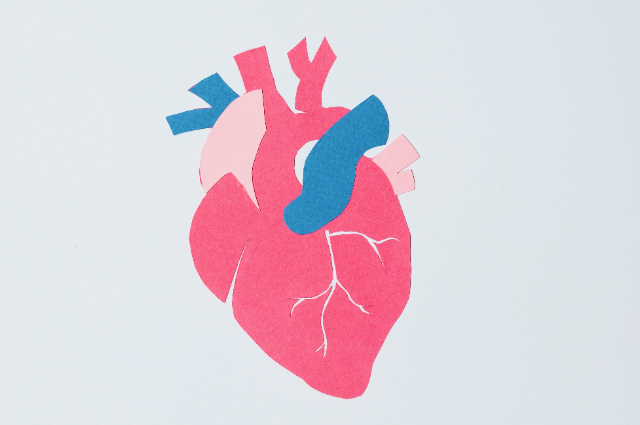
To understand what goes wrong in aortic stenosis, it helps to picture how a healthy aortic valve is built. The heart has four chambers and four valves. Blood returning from the body enters the right side, is sent through the lungs to collect oxygen, and returns to the left side. From the left atrium, it passes through the mitral valve into the left ventricle, which then contracts and drives the blood out through the aortic valve into the aorta, the great artery from which every other artery in the body ultimately branches. The aortic valve is therefore the final gate that the blood must cross before it is delivered to the brain, the kidneys, the muscles and the heart itself.
A normal aortic valve has three thin, flexible leaflets, called cusps, shaped somewhat like small half-moons or pockets. They are attached to a ring of tissue at the base of the aorta, known as the annulus, and each cusp sits above a small bulge in the aortic wall called a sinus. When the ventricle contracts, pressure pushes the three cusps flat against the walls of the aorta and the valve opens fully, leaving an area of roughly three to four square centimetres through which blood can rush. When the ventricle relaxes, blood tries to fall back, fills the pockets, and forces the three cusps to meet in the centre and seal the opening.
This closure is what keeps the blood moving forward and gives the heart time to refill. Two of the sinuses give rise to the coronary arteries, the small vessels that feed the heart muscle. This detail, which might seem a curiosity, turns out to be enormously important in modern practice. Whenever a replacement valve is placed inside a native valve, the old diseased leaflets are pushed aside, and the surgeon or cardiologist must be certain that they do not block the openings of the coronary arteries. The distance between the annulus and the coronary openings, and the width of the sinuses, are measured carefully before every procedure for exactly this reason.
The valve works under remarkable conditions. It opens and closes more than thirty million times a year, and each closure must withstand the pressure of the blood in the aorta, which in a healthy adult is often around one hundred millimetres of mercury and can be much higher in a person with hypertension. The leaflets are made of layers of collagen, elastic fibres and specialised cells, arranged so that they can bend easily and yet bear heavy loads. They are also alive, in the sense that they contain cells capable of sensing mechanical stress and repairing minor damage. This is why a healthy valve can serve for a lifetime.
Not everyone is born with the standard design. About one or two people in every hundred are born with a bicuspid aortic valve, in which two of the three cusps have fused during development so that only two leaflets are present. A bicuspid valve may function perfectly well for decades, but its geometry exposes the leaflets to abnormal stresses, and it tends to calcify and narrow ten to fifteen years earlier than a normal valve. Many people with a bicuspid valve also have a dilated aorta, which adds a further layer of complexity when treatment is being planned.
Knowing the anatomy also explains why the valve cannot simply be repaired in most cases of stenosis. The leaflets are not merely dirty or scarred on the surface. They are thickened, hardened and fused, and the calcium is embedded in the tissue itself. Attempts to break the stiffness by inflating a balloon inside the valve, a procedure known as balloon valvuloplasty, give only a brief and unreliable improvement. The valve, once it has hardened, needs to be replaced, and it is that need which has driven a century of surgical ingenuity and, more recently, the invention of the transcatheter approach.
When the Door Stiffens: How Aortic Stenosis Develops
For a long time, calcific aortic stenosis was explained as simple wear and tear, a mechanical consequence of decades of opening and closing, like the hinge of a gate that eventually rusts. Research over the past thirty years has shown that the truth is more interesting. The process resembles, in several respects, the biology of atherosclerosis, the same disease that clogs the coronary arteries. The valve surface is subjected to constant mechanical stress, particularly on the side facing the aorta, and small injuries to the lining allow lipids and inflammatory cells to enter the tissue. Over years, this sets off a chain of reactions in which cells in the leaflet change their character, begin to behave like bone-forming cells, and lay down deposits of calcium.
The result is a valve that gradually becomes thicker and less flexible. In the earliest stage, called aortic sclerosis, the leaflets are thickened and slightly calcified, but the opening is still wide enough for blood to pass freely. Sclerosis is very common in older adults, and in most people it advances slowly or not at all. In a smaller group, however, the process accelerates. The calcium deposits enlarge, the leaflets stiffen and begin to fuse at their edges, and the opening narrows. Once the narrowing becomes significant, the term stenosis is applied, and the burden on the heart begins to grow.
A number of factors influence who is affected and how fast the disease advances. Age is the strongest of them. Studies that combined data from several countries have estimated that a little over twelve in every hundred people aged seventy-five or older have some degree of aortic stenosis, and that around three or four in every hundred have the severe form. Male sex, high blood pressure, raised cholesterol, diabetes, smoking and chronic kidney disease all appear to speed the process. A bicuspid valve, as noted earlier, brings the disease forward by more than a decade. Some families seem to carry a higher risk, which suggests that genetic factors also play a part.
Not all stenosis is calcific. In many parts of the world, and especially in low-income and middle-income countries, rheumatic heart disease remains an important cause of valve damage. Rheumatic fever, which follows an untreated streptococcal throat infection in childhood, can trigger an immune reaction that scars the heart valves. The aortic valve is affected less often than the mitral valve, but when it is, the scarring tends to fuse the edges of the leaflets, producing a narrowed and often leaky valve in a much younger person. Global estimates suggest that rheumatic heart disease still affects tens of millions of people, and India carries a substantial share of that burden. This difference matters, because the treatments that suit an elderly patient with calcific stenosis do not always suit a younger patient with rheumatic damage.
What happens to the heart while the valve narrows is a story of adaptation followed by failure. As the opening shrinks, the left ventricle must generate much higher pressure to eject the same volume of blood. The muscle responds by growing thicker, a change called hypertrophy, which initially helps by reducing the stress on each muscle fibre. But a thick muscle is also a stiff muscle. It relaxes poorly, it needs more oxygen than the coronary arteries can readily supply, and it becomes prone to scarring and to rhythm disturbances. Over the years, the compensation that once protected the heart begins to work against it. Cardiologists grade the severity of stenosis using measurements taken during echocardiography. In severe stenosis, blood crosses the valve at a velocity of four metres per second or more, the average pressure difference across the valve is at least forty millimetres of mercury, and the valve area falls to about one square centimetre or less, compared with three or four in a normal valve. These numbers are not arbitrary. They mark the point at which the risk of symptoms, heart failure and death begins to climb steeply.
The most important feature of the natural history of aortic stenosis is its two-phase character. For years, sometimes for decades, the disease is silent, and the outlook is good. Once symptoms appear, the picture reverses. A classical description from the late 1960s estimated that the average survival after the onset of chest pain was about five years, after fainting about three years, and after heart failure about two years. Modern series show that a large proportion of patients with untreated symptomatic severe stenosis die within a couple of years. No medicine has been shown to reverse the process, which is why replacement of the valve, and not medical therapy, is the treatment that changes the outcome.
Whispers Before the Storm: Symptoms and Their Meaning
The difficulty with aortic stenosis is not that its symptoms are dramatic. It is that they are so easy to mistake for something else. In the early years, there may be nothing to notice at all. Later, a person who used to walk to the market without stopping may begin to pause halfway. A grandmother who climbed the stairs of her home several times a day may start to avoid them. A man who worked in the garden until dusk may find that he has to sit down after an hour. Because these changes happen slowly and because they coincide with growing older, families and even patients themselves often explain them away. It is only when the symptoms become impossible to ignore that medical help is sought.
The classical triad of symptoms in aortic stenosis consists of breathlessness, chest discomfort and fainting. Breathlessness on exertion is the most common. It arises because the stiff, thickened ventricle fills at a higher pressure, which is transmitted backwards into the lungs and makes breathing feel laboured. In the later stages, breathlessness may occur at rest or on lying flat, and the person may need extra pillows to sleep. Chest discomfort, which resembles the angina of coronary artery disease, occurs because the enlarged muscle needs more blood than the coronary arteries can deliver, even when those arteries are clear. It is typically brought on by effort and relieved by rest.
Fainting, or near-fainting, is the third and often the most alarming feature. During exertion, the muscles of the body open their blood vessels wide to receive more blood, but a narrowed valve limits how much the heart can push out. Blood pressure may fall abruptly and the brain is briefly starved of supply. A person who blacks out while climbing a hill, carrying a load or hurrying to catch a bus should be examined promptly. Dizziness on standing or exertion deserves the same attention. In some cases, rhythm disturbances of the heart contribute to these episodes.
Other, less specific symptoms often appear. Extreme tiredness, reduced tolerance for activity, a feeling of heaviness in the chest, palpitations and swelling of the ankles may all occur as heart failure develops. Some older people, and especially those who have already reduced their activity to avoid discomfort, may report very little. Their disease can advance quietly behind a life that has slowly shrunk to fit the limits of the heart. Physicians therefore ask direct questions about what a patient has stopped doing over the past year, because the answer is often more revealing than a list of complaints.
A doctor listening to the chest may detect a harsh murmur, best heard over the upper right side of the breastbone and often radiating toward the neck. This sound is produced by turbulent blood crossing the narrowed valve, and it usually becomes louder as the stenosis worsens, until in very severe cases the flow is so restricted that the murmur becomes softer again. A weak, slow-rising pulse and a narrow pulse pressure are further clues. In many older patients, a murmur is first noticed during a routine examination, long before any symptoms occur, which is one reason why regular check-ups remain valuable.
Why does the appearance of symptoms matter so much? Because it changes the decision. In a person with severe stenosis and no symptoms, doctors may follow the patient closely and time the intervention carefully. Once symptoms develop, delay becomes hazardous, and the guidelines of major cardiology societies recommend that valve replacement be considered without undue waiting. The message for the public is straightforward. Breathlessness, chest discomfort or fainting in an older person should not be accepted as normal ageing until the heart valve has been checked, particularly if a murmur has ever been mentioned.
Listening with Sound Waves: Diagnosis and Imaging
The diagnosis of aortic stenosis begins with a conversation and a stethoscope, but it is confirmed by imaging. The central test is the transthoracic echocardiogram, a painless examination in which a small probe placed on the chest sends out high-frequency sound waves and records the echoes that return from the heart. The machine converts these echoes into moving pictures, so the doctor can watch the valve leaflets open and close in real time, see how thick and calcified they are, and observe the size and strength of the ventricle. It involves no radiation, needs no injection in most cases, and can be repeated as often as needed.
Beyond simple pictures, echocardiography uses the Doppler principle to measure the speed of blood as it crosses the valve. The faster the flow, the tighter the narrowing. From that velocity, the machine can calculate the pressure difference across the valve, and by combining it with measurements of the outflow tract, it can estimate the valve area. Together, these figures allow the doctor to classify the disease as mild, moderate or severe, and to decide how often the patient should be reviewed. In mild disease, an interval of several years may be acceptable, whereas in severe disease, follow-up may be needed every six to twelve months.
Some patients are more difficult to classify. In a group sometimes described as having low flow, low-gradient stenosis, the valve area looks severely reduced, but the pressure difference is lower than expected because the ventricle is too weak, or too small and stiff, to push out a normal volume of blood. Such patients may still have truly severe stenosis and may benefit from replacement, but proving it requires additional testing. A low-dose dobutamine stress echocardiogram can show whether the heart muscle has reserve, and a computed tomography scan can measure the calcium in the valve directly. A high calcium score, with thresholds that differ for men and women, strongly supports a diagnosis of severe disease.
Computed tomography has become the second pillar of diagnosis, particularly when a transcatheter procedure is being considered. A specially timed scan of the heart and of the arteries from chest to groin produces a detailed three-dimensional map. It allows the team to measure the size and shape of the aortic annulus with millimetre accuracy, to judge how much calcium is present and where it lies, to check how far the coronary arteries sit from the valve, and to assess whether the vessels in the abdomen and legs are wide enough to carry the catheter. A valve chosen without this information would be a guess; a valve chosen with it is a considered decision.
Other tests complete the picture. A coronary angiogram, in which dye is injected into the arteries of the heart, is usually performed to look for blockages, because many older patients with stenosis also have coronary disease and the treatment plan may need to address both. Blood tests assess kidney function, blood count and markers of heart strain. An electrocardiogram records the rhythm and shows whether there are conduction problems that may influence the choice of valve. A frailty assessment, which examines strength, walking speed, nutrition and mental function, has also become an essential part of decision-making.
All these tests serve a single purpose: to determine how severe the disease is, how the heart has responded, and which form of treatment gives this particular person the best chance of a long and comfortable life. In modern practice, the results are not interpreted by a single doctor in isolation. They are presented to a multidisciplinary group, the heart team, whose role is discussed later. The value of accurate imaging, in the end, lies not in the pictures themselves but in the decisions that they make possible.
From Scalpel to Catheter: The Road to a New Treatment
The story of aortic valve replacement begins in the middle of the twentieth century. In the early 1960s, surgeons in the United States and Britain implanted the first artificial valves in the aortic position, using a ball-and-cage design in which a small metal or silicone sphere moved inside a cage to open and close the passage. These devices were crude by present standards, but they saved lives that would otherwise have been lost. Over the decades that followed, engineers and surgeons refined the designs, introducing tilting-disc and bileaflet mechanical valves, and later biological valves made from animal tissue mounted on a supporting frame. Surgical aortic valve replacement became one of the most reliable operations in heart surgery.
Yet the operation retained its essential demands. It required a cut through the breastbone, the use of a heart-lung machine, and a period during which the heart was deliberately stopped. In a healthy sixty-year-old, these are manageable stresses. In an eighty-five-year-old with weak lungs, kidney disease and a history of stroke, they can be overwhelming. Surveys conducted in Europe in the early 2000s found that a substantial fraction of elderly patients with severe symptomatic stenosis were never offered surgery at all, because they were judged too risky. The disease was clearly treatable, but the treatment was out of reach for the very people who most often suffered from it.
The intellectual seeds of a less invasive method were planted in the 1980s and 1990s, when interventional cardiologists learned to open blocked coronary arteries by threading balloons and stents through catheters. It seemed reasonable to ask whether a valve could be delivered in the same way. Early experiments with balloon valvuloplasty in the 1980s showed that stretching a stenotic valve could give temporary relief, but the benefit faded within months as the valve narrowed again. Several researchers, including Danish and American investigators, proposed stent-mounted valves during the same period, but the idea remained largely theoretical until it could be tested in patients.
That test came on the sixteenth of April 2002, in the French city of Rouen. A cardiologist named Alain Cribier, who had spent years developing a balloon-expandable valve made from bovine pericardial tissue sewn inside a stainless-steel stent, implanted it in a man in his late fifties who was in cardiogenic shock and considered too ill for surgery. The valve was delivered through a vein and across the wall between the two upper chambers of the heart, and it was expanded within the narrowed native valve. The patient improved dramatically. The report published later that year in the journal Circulation described the first human implantation of a catheter-delivered aortic valve, and it is generally regarded as the starting point of the modern era. Progress over the next decade was steady, if cautious. Early devices were large and hard to steer, and the first patients were extremely sick. Access routes were refined, with the artery of the leg soon replacing the vein as the preferred entry point. Two principal designs emerged. One used a balloon to expand a stent-mounted valve; the other used a self-expanding frame made of a nickel and titanium alloy that opened by itself when released from its sheath. Each had its own strengths, and both continue to be improved. Companies, hospitals and national regulators began the long task of testing the new technology in formal trials.
The trials transformed the field. In 2010, a landmark randomised study called PARTNER, in its cohort of patients who could not undergo surgery, showed that TAVI reduced deaths at one year from about fifty-one in every hundred to about thirty-one in every hundred compared with standard medical treatment. A companion study the next year, in patients who were at high risk but still operable, found that TAVI and surgery gave similar survival at one year. Later trials extended the comparison to patients of intermediate surgical risk, and then, in 2019, to those at low risk. The results, published in the New England Journal of Medicine, showed outcomes with TAVI that were at least comparable to surgery in the periods studied.
These results changed practice guidelines across the world. Cardiology societies in Europe and North America now recommend TAVI as the preferred option for older patients, with the age threshold set at about seventy-five in Europe and eighty in the American guidance, and shared decision-making advised in the range between about sixty-five and eighty. A procedure that started as a rescue for the hopeless is now, in many centres, the commonest way of replacing the aortic valve. The transition from experiment to standard care in barely two decades is among the fastest in the history of cardiac medicine, and it reflects both the ingenuity of the engineers and the discipline of the clinical trials that tested them.
Through the Groin to the Heart: How a TAVI Procedure Works
A TAVI procedure is carried out in a specially equipped room, often called a hybrid operating theatre or catheterisation laboratory, which combines the imaging systems of a cardiology suite with the sterility and emergency capability of a surgical operating room. The team typically includes an interventional cardiologist, a cardiac surgeon, an anaesthetist, an imaging specialist, nurses and technicians. Their presence reflects the fact that, although the procedure is minimally invasive, it operates on one of the most important structures in the body, and complications must be handled without delay.
Preparation begins long before the patient reaches the theatre. The computed tomography scan described earlier is used to choose the type and size of the valve. A valve that is too small may leak around its edges or may slip; one that is too large may injure the annulus or rupture the surrounding tissue. The planners also decide on the route of entry. In more than nine of every ten cases in modern practice, the chosen route is transfemoral, meaning that the catheter is inserted through the femoral artery at the top of the thigh. If the leg arteries are too narrow, diseased or twisted, other routes may be used, including the arteries of the shoulder or neck, or a small incision between the ribs.
On the day of the procedure, many patients receive only local anaesthetic at the puncture site together with light sedation, and remain awake and able to speak to the team. General anaesthesia is used in some patients, particularly if complex imaging or difficult access is expected. A small tube is placed in the femoral artery, and through it the operator passes a thin wire, which is guided under continuous X-ray imaging across the narrowed valve and into the left ventricle. This wire acts as a rail along which all other devices will travel. A temporary pacing wire is usually positioned in the right ventricle, because the heart will be briefly driven at a very rapid rate during valve deployment.
The replacement valve, crimped to a small size and mounted on a delivery catheter, is then advanced along the wire, through the aorta and into the position of the old valve. The operator confirms the position using X-ray fluoroscopy and, in some centres, echocardiography. The moment of deployment requires precision. With a balloon
expandable valve, rapid pacing is started to reduce the movement of the heart and the balloon is inflated, pressing the valve against the old leaflets and the annulus. With a self-expanding valve, the outer sheath is slowly withdrawn, and the frame opens under its own spring force, sometimes with the option of recapturing and repositioning it before the final release.
Once the valve is in place, its function is checked at once. The operator injects contrast dye to see whether blood is leaking backwards, measures the pressure difference across the new valve, and uses ultrasound to confirm smooth opening and closing. If a small leak is found around the edge of the valve, a gentle additional balloon inflation may correct it. When the team is satisfied, the catheters are withdrawn, and the puncture in the artery is closed, either with a stitching device placed beforehand or with a small closure plug. The entire procedure, from puncture to closure, commonly lasts between one and two hours.
The new valve works from the moment it opens. Blood that had been struggling through an opening of one square centimetre is suddenly able to flow through a passage two or three times as wide, and the pressure inside the ventricle falls almost immediately. Patients often report that they feel the difference in their breathing within days. Behind that simple experience lies a remarkable convergence of imaging, materials science and technical skill, and it is because so many of the steps can be rehearsed and standardised that the procedure has become a routine option rather than a heroic one.
Choosing the Right Path: Patient Selection and the Heart Team
No treatment suits everyone, and TAVI is no exception. The claim that it has replaced surgery is a simplification, because the choice between the two remains a matter of individual judgment. The decision depends on a large number of features of the patient and of the valve, and it is best made not by a single physician but by a group. This group, known as the heart team, brings together an interventional cardiologist, a cardiac surgeon, an imaging specialist, an anaesthetist and, often, a geriatrician or nurse coordinator. Its central purpose is to ensure that the recommendation reflects several viewpoints and that the patient receives the treatment most likely to serve him or her over the whole of life.
Age is the first and most visible factor, but it is used as a guide rather than a rule. For older patients, generally those beyond the late seventies, the shorter life expectancy means that the durability of the valve is less critical, and the gentler nature of TAVI offers a clear advantage. For younger patients, the equation changes. A person in their fifties or sixties may live for another thirty years, and the valve must be expected to last. Because mechanical valves, which are highly durable, need lifelong treatment with blood-thinning medication, and because tissue valves gradually wear out, the choice for a younger person involves a long-term strategy and not a single operation.
Other factors carry great weight. General health, including the state of the lungs, kidneys and brain, affects the ability to tolerate a major operation. Frailty, assessed through walking speed, grip strength and independence in daily activity, has proved to be a powerful predictor of outcome. Anatomical considerations decide whether the transcatheter route is technically possible: a very small or very large annulus, heavily calcified leaflets that might injure the coronary openings, a bicuspid valve, a dilated aorta, or severe tortuosity of the leg arteries can all favour surgery. The presence of coronary artery disease that requires bypass grafts, or of another diseased valve that needs simultaneous repair, may also point toward an open operation.
The condition of the valve itself is relevant as well. In rheumatic disease, where the leaflets are scarred and often accompanied by mitral valve involvement, surgery is generally preferred. Bicuspid valves, which are typically heavily calcified and asymmetrical, pose a higher risk of poor expansion or leakage and are usually judged case by case. A valve that is already failing because of infection cannot be treated by TAVI in the standard manner. These exceptions show why the guidelines speak of recommendations and not of prescriptions.
One of the most important considerations is the lifetime plan. A tissue valve, whether surgical or transcatheter, has a limited lifespan, generally estimated at ten to fifteen years for surgical bioprostheses and still being defined for transcatheter valves. A person who receives a TAVI valve at seventy may never need another intervention, but one who receives it at sixty-five may. The first valve should therefore be chosen in a way that keeps later options open, including the possibility of placing a second transcatheter valve inside the first, a technique called valve-in-valve. The design of the first implant may make the second easier or harder, especially with respect to future access to the coronary arteries.
Finally, the patient's own values must be heard. Some people place the highest priority on a short hospital stay and a quick return to independence. Others prefer the long track record of surgery and are willing to accept a longer recovery in exchange. Good decision-making means explaining the benefits, the risks and the uncertainties in plain language, and letting the patient and family take part. A recommendation that is technically excellent but ignores what the patient wants from the rest of life has not fully done its job.
Recovery and Return to Life: After the Procedure
One of the most striking features of TAVI is how quickly most patients recover. After the procedure, the patient is usually monitored for several hours in a recovery area or a cardiac care unit, where the heart rhythm, blood pressure and the puncture site in the groin are watched closely. Because no incision is made in the chest, there is no breastbone to heal and far less pain. Most patients are encouraged to sit up and take a few steps within a day. In uncomplicated cases, discharge from hospital is possible within two or three days, compared with a week or more after open surgery.
Before leaving hospital, the patient usually undergoes an echocardiogram to confirm that the new valve is working well, and an electrocardiogram to look for rhythm problems. The heart's electrical wiring runs close to the aortic valve, and pressure from the new valve can sometimes disturb it. A minority of patients develop a conduction block that requires a permanent pacemaker, and this is one of the reasons why monitoring after the procedure matters. Medication is adjusted as well. Most patients receive an antiplatelet drug, and those who already need anticoagulants for another condition, such as atrial fibrillation, continue with them under specialist guidance.
The recovery at home is usually straightforward but should not be casual. The puncture site should be watched for swelling, bleeding or increasing pain. Gentle walking is encouraged from the first days, and most patients return to light domestic activity within a week, though heavy lifting and strenuous exercise are usually delayed for a few weeks. Many centres offer a programme of supervised exercise, sometimes called cardiac rehabilitation, which helps older patients regain muscle strength that may have been lost during months of restricted activity before the procedure. Good nutrition and adequate sleep contribute more to recovery than is often realised.
Patients and families frequently ask whether there is any difference in dental care or infection risk. There is. An artificial valve, whether surgical or transcatheter, carries a small risk of infective endocarditis, an infection of the valve that can be serious. For this reason, good oral hygiene and regular dental review are strongly recommended, and antibiotics before certain dental procedures are advised for patients with prosthetic valves. Any unexplained fever that lasts more than a day or two, especially after a dental or skin procedure, should prompt a medical consultation and blood cultures before antibiotics are started.
The benefits for daily life can be considerable. In clinical studies, patients report clear improvement in breathlessness, walking distance and general well-being within weeks of the procedure, and quality-of-life scores climb sharply in the first month. For many older people, the change is not merely physical. Being able to walk to a place of worship, to attend a wedding, to play with grandchildren or to travel again restores a sense of independence that the disease had quietly stripped away. Stories of very elderly patients who were breathless on the eve of the procedure and were out visiting neighbours within the week show what the technology can do at its best, although individual outcomes naturally vary.
Follow-up is lifelong. Patients are usually reviewed at one month, again at about one year, and then at regular intervals, with echocardiography to detect any change in valve function. The purpose is not to look for trouble but to catch it early if it does arise. A tissue valve does not last forever, and the reassuring outcome of the first few years should not be mistaken for permanent cure. Regular review, control of blood pressure and cholesterol, avoidance of tobacco and attention to other heart conditions all contribute to keeping the valve, and the person who carries it, in good health for as long as possible.
Risks, Limits and the Question of Durability
Every treatment carries risks, and an honest account of TAVI must give them equal attention. The procedure is less invasive than surgery, but it is not free of danger. Serious complications are uncommon in experienced centres but can be devastating when they occur. Stroke is one of the best known. During valve deployment, fragments of calcium or clot can break free from the diseased leaflets and travel to the brain. The rate of disabling stroke in recent trials has been low, generally in the range of one or two in every hundred patients, but it is not zero, and protection devices that catch debris are under continued study.
Damage to the blood vessels at the access site is another possibility. The catheters used for TAVI are much smaller than they once were, and modern closure devices have reduced bleeding, but tears, blockages or bleeding in the groin still occur in a small proportion of patients. Kidney injury may follow the contrast dye used for imaging, particularly in patients whose kidneys were already weak. Rarely, the annulus or the aorta may rupture during expansion of the valve, an event that requires emergency surgery, which is one reason why TAVI programmes must have surgical backup readily available.
Rhythm disturbances are among the commonest complications. Because the conduction system of the heart lies beside the aortic valve, compression by the new implant may cause a block in electrical signals. The proportion of patients needing a pacemaker after TAVI has varied between devices and techniques, with self-expanding valves generally producing higher rates than balloon-expandable ones, and it is an important consideration for younger patients who will live with the pacemaker for many years. Refinements in technique, including higher and more careful positioning of the valve, have reduced this risk over time.
A further issue is leakage around the valve, known as paravalvular regurgitation. Because a transcatheter valve is pressed into a calcified and irregular annulus, small gaps may remain between the frame and the tissue through which blood leaks backwards. Early devices were prone to moderate or severe leaks, which were linked to poorer survival. Newer designs, with an outer skirt or sealing cuff and improved sizing based on computed tomography, have made significant leaks rare, but they remain a concern in heavily calcified or bicuspid valves.
The most discussed limit of TAVI is durability. Surgical bioprostheses have been followed for decades, and their behaviour is reasonably well understood, with structural deterioration becoming increasingly common after ten to fifteen years. Transcatheter valves are far newer. Trials and registries now provide follow-up extending to five years and beyond in large groups and to about ten years in smaller ones, and so far the results suggest that the valves perform well and that the rate of structural failure is low. But the patients in these studies were mostly elderly, and many died of other causes before a valve could fail. The evidence for patients who receive a valve at sixty or sixty-five and live for another thirty years is simply not yet available.
This uncertainty explains the caution of the guidelines for younger patients and the emphasis on lifetime management. It also explains active research on newer materials, on treatments that reduce calcification of tissue valves and on the technique of implanting a second valve within a failing first one. Honest communication about what is not yet known is part of good medical practice. The best available evidence is encouraging, but it is still incomplete, and a patient who understands this can make a decision that he or she will not regret.
Looking Ahead: Technology, Access and Equity
The future of aortic valve treatment is being shaped by several parallel developments. On the technological side, valve designs continue to improve, with lower profiles for easier delivery, better sealing, more flexible frames that allow future access to the coronary arteries, and leaflet materials chosen to slow wear. Embolic protection devices, better imaging, and artificial intelligence tools that help analyse computed tomography scans are being studied to make the procedure safer and more predictable. Research is also examining whether earlier intervention in patients with severe stenosis who have no symptoms, or with only moderate disease, can prevent irreversible damage to the heart muscle.
At the same time, the population that needs treatment is growing. As life expectancy rises in most parts of the world, the number of people living into their eighties and nineties increases, and with it the number who will develop calcific aortic stenosis. Some estimates suggest that the number of patients eligible for valve replacement in developed countries will roughly double over the coming decades. Health systems will need to plan for this demand, not only by providing procedure rooms and devices but also by training the imaging specialists, nurses and surgeons on whom the heart team depends.
For a country such as India, the challenge is different in kind and in scale. The population is large and ageing, the burden of rheumatic heart disease remains substantial, and access to specialised cardiac care is uneven, with advanced centres concentrated in large cities. The cost of imported valves has been a major barrier, since a single device may cost more than the annual income of many families. The public insurance schemes and state programmes that pay for cardiac surgery do not always cover transcatheter valves, and patients in rural areas may have to travel hundreds of kilometres for assessment. These circumstances mean that many people who could benefit from TAVI do not have any realistic access to it.
Efforts to close this gap are under way. Several manufacturers in India and other emerging economies have developed indigenous transcatheter valves, some of which have been tested in clinical studies and approved for use, and their prices are lower than those of imported devices. Training programmes for heart teams, tele-consultation between district hospitals and tertiary centres, and inclusion of valve procedures in public insurance packages could all extend access. None of these solutions is simple, and each requires sustained investment and honest evaluation. The aim should be to ensure that a decision about treatment is made on the basis of medical need and not on the basis of postal code or income.
Prevention and early detection deserve equal attention. Rheumatic heart disease is largely preventable through prompt treatment of streptococcal throat infections and secondary prophylaxis with penicillin in those who have had rheumatic fever. Control of blood pressure, cholesterol, diabetes and tobacco use may slow the progression of calcific disease, although no drug has yet been proved to halt it. A simple stethoscope examination by a trained primary care doctor, followed by an echocardiogram when a murmur is heard, remains one of the most cost-effective ways of finding valve disease before it becomes an emergency.
The future of valve treatment, therefore, is not only a technological story. It is also a story of public health, medical education and social policy. The catheter-based valve is an achievement of engineering, but its value depends on how widely and fairly it can be offered. The measure of success will be whether the benefits reach the many ordinary patients who need them, rather than a fortunate few who happen to live near the right hospital.
The story of the aortic valve is, in a sense, the story of modern cardiology in miniature. A structure that for centuries was a source of silent suffering has become one that can be understood, measured and replaced. The valve narrows through a slow biological process linked to age, inflammation and calcium; the heart adapts and then fails; and once symptoms begin, the outlook without treatment is poor. Recognising that pattern, and acting on it promptly, remains the single most important step in the care of a patient with aortic stenosis.
Transcatheter aortic valve implantation has altered the options open to that patient. In less than twenty-five years, it has moved from a single daring implantation in Rouen to a widely used treatment supported by large randomised trials, and it has given older and frailer patients a chance of relief that once was denied to them. The procedure reduces the physical burden of treatment, shortens hospital stay and allows a rapid return to daily life. For those who are suitable, it can restore both years and the quality of those years.
At the same time, the technology should be viewed with balanced judgment. It is not appropriate for everyone, and surgery remains the better choice in many situations, including rheumatic disease, certain bicuspid valves, and cases where other heart operations are needed at the same time. Complications such as stroke, rhythm disturbance and valve leakage still occur, and the long-term durability of transcatheter valves in younger patients is not yet known. These limits do not diminish the achievement; they explain why the decision must be made carefully, by a heart team, in partnership with the patient and family.
The wider lessons are equally important. Symptoms such as breathlessness, chest discomfort and fainting in an older person must never be dismissed as simple ageing. A murmur discovered during a routine check deserves an echocardiogram. Prevention of rheumatic fever and control of common risk factors deserve continued public health investment. And societies must ask how the benefits of costly technology can be extended to the people who need them, whether they live in a metropolitan hospital district or a distant village.
A final point concerns humility. Medicine has a habit of celebrating each new tool as though it were the last word, and later evidence sometimes corrects early enthusiasm. The sensible response to a promising technology is neither blind faith nor reflex suspicion but continued measurement: long registries, honest reporting of complications, and willingness to change advice when the data change. If the field keeps to that discipline, the coming decades of follow-up will show how far the early promise of TAVI can be extended, and patients will be better served for it.
On World Heart Day, it is worth remembering that progress in cardiology is not measured only in devices and trials. It is measured in the grandmother who can walk to the temple again, the farmer who can return to his fields, and the retired clerk who can travel to see his family again. The aortic valve is a small door, but what passes through it is life itself. Understanding how to keep that door open is one of the quiet triumphs of modern medicine, and the task now is to ensure that this knowledge reaches everyone who needs it.
References:
- Adams DH, Popma JJ, Reardon MJ, et al. Transcatheter aortic-valve replacement with aself-expanding prosthesis. N Engl J Med. 2014;370:1790-1798.
- Baumgartner H, Hung J, Bermejo J, et al. Recommendations on the echocardiographic assessment of aortic valve stenosis: a focused update from the European Association of Cardiovascular Imaging and the American Society of Echocardiography. J Am Soc Echocardiogr. 2017;30:372-392.
- Carabello BA, Paulus WJ. Aortic stenosis. Lancet. 2009;373:956-966.
- Coffey S, Roberts-Thomson R, Brown A, et al. Global epidemiology of valvular heart disease. Nat Rev Cardiol. 2021;18:853-864.
- Cribier A, Eltchaninoff H, Bash A, et al. Percutaneous transcatheter implantation of an aortic valve prosthesis for calcific aortic stenosis: first human case description. Circulation. 2002;106(24):3006-3008.
- Leon MB, Smith CR, Mack M, et al. Transcatheter aortic-valve implantation for aortic stenosis in patients who cannot undergo surgery. N Engl J Med. 2010;363:1597-1607.
- Leon MB, Smith CR, Mack MJ, et al. Transcatheter or surgical aortic-valve replacement in intermediate-risk patients. N Engl J Med. 2016;374:1609-1620.
- Mack MJ, Leon MB, Thourani VH, et al. Transcatheter aortic-valve replacement with aballoon-expandable valve in low-risk patients. N Engl J Med. 2019;380:1695-1705.
- Osnabrugge RLJ, Mylotte D, Head SJ, et al. Aortic stenosis in the elderly: disease prevalence and number of candidates for transcatheter aortic valve replacement. J Am Coll Cardiol. 2013;62:1002-1012.
- Otto CM, Nishimura RA, Bonow RO, et al. 2020 ACC/AHA guideline for the management of patients with valvular heart disease. Circulation. 2021;143:e72-e227.
- Popma JJ, Deeb GM, Yakubov SJ, et al. Transcatheter aortic-valve replacement with aself-expanding valve in low-risk patients. N Engl J Med. 2019;380:1706-1715.
- Ross J Jr, Braunwald E. Aortic stenosis. Circulation. 1968;38(1 Suppl):61-67.
- Smith CR, Leon MB, Mack MJ, et al. Transcatheter versus surgical aortic valve replacement in high-risk patients. N Engl J Med. 2011;364:2187-2198.
- Vahanian A, Beyersdorf F, Praz F, et al. 2021 ESC/EACTS guidelines for the management of valvular heart disease. Eur Heart J. 2022;43:561-632.
- Watkins DA, Johnson CO, Colquhoun SM, et al. Global, regional, and national burden of rheumatic heart disease, 1990-2015. N Engl J Med. 2017;377:713-722.