Image by Colin Behrens from Pixabay
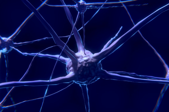
Our bodies move due to a complex system of signals travelling from the brain to our muscles. However, these signals don't always take a direct route. They often pass through a network of "switchboard operator" cells within the spinal cord called interneurons. These interneurons play a crucial role in shaping and refining the signals that ultimately control our movements.
A New Map for Navigating the Motor System
To better understand this complex network, researchers at St. Jude Children's Research Hospital created a comprehensive map of the brain. This map highlights the specific brain regions that directly connect to a particular type of interneuron known as V1 interneurons. These V1 interneurons are essential for proper movement. This groundbreaking map along with an interactive 3D website has provided a valuable resource for scientists to explore the complex connections within the nervous system. It offers a clearer picture of how different parts of the brain communicate with the spinal cord to generate movement.
The Challenge of Studying Interneurons
While significant progress has been made in understanding how different brain regions contribute to motor control, highlighting the exact connections between these regions and specific spinal cord neurons has remained a major challenge. The difficulty lies in the sheer diversity of interneurons. These cells come in hundreds of different types, all intricately intertwined. Studying these diverse cell types individually has been a significant hurdle for researchers.
The Significance of this Research
This new brain map represents a significant step forward in our understanding of how the brain controls movement. By providing a clearer picture of the connections between the brain and the spinal cord, this research will pave the way for future studies that delve deeper into the mechanisms underlying motor function. This knowledge has the potential to shed light on various neurological conditions that affect movement such as spinal cord injuries and neurodegenerative diseases. By understanding the complex workings of the motor system, researchers can develop more effective treatments and therapies for these conditions.
The development of this comprehensive brain map is a testament to the ongoing efforts to unravel the mysteries of the nervous system. By providing a clearer understanding of the complex connections within the motor network, this research will undoubtedly have a profound impact on our understanding of movement and its underlying neurological mechanisms.
Untangling Neural Communication: A Groundbreaking Look at Brain-Spinal Cord Connections
The intricate network of neural communication within the body can be a challenge. However, this task is far more complex as it involves decoding signals shaped by over three billion years of evolution. Dr. Anand Kulkarni, one of the lead researchers had described it as a task of extraordinary complexity that required meticulous methods to uncover the mechanisms of neural interaction.
Exploring Interneuron Subclasses
Recent advancements in neuroscience have shed light on the existence of distinct interneuron subclasses both molecularly and developmentally. Despite this progress, many questions remain unanswered, particularly about how these subclasses fit into the larger framework of neural communication. As Dr. Bikoff explains, understanding how descending motor systems connect to cellular targets is essential to uncovering the brain's methods of controlling movement and behaviour. This knowledge could help scientists decipher the pathways through which the brain sends and receives signals.
A Novel Approach Using Modified Viruses
To unravel the circuits connecting the brain to the spinal cord, researchers employed an innovative method involving a genetically modified rabies virus. Normally, this virus spreads between neurons by utilizing a surface protein called glycoprotein. However, by removing the glycoprotein, researchers prevented the virus from travelling beyond its initial location.
To delve deeper, they reintroduced the glycoprotein to a specific group of interneurons. This modification allowed the virus to make a single leap across synapses after which it became stuck again. By tagging the virus with a fluorescent marker, scientists could track its movement and determine the regions of the brain connected to these interneurons.
Mapping Neural Connections in 3D
Using this advanced technique, researchers focused on V1 interneurons as a class known for their crucial role in shaping motor output. This approach enabled them to create a detailed 3D map of neural connections by accurately tracing the pathways of various signals back to the brain.
These findings represent a significant step forward in visualizing and understanding the intricate connections between the brain and spinal cord. By revealing these pathways, the study provides new insights into how neural signals coordinate movement and behaviour, opening doors to further exploration in neuroscience. Through innovative methods and detailed mapping, this research illuminates the complexities of neural communication, bringing us closer to understanding the intricate design of the human nervous system.
Illuminating Neural Pathways: Tracking Brain-to-Spinal Cord Connections
Understanding the brain's intricate communication with the spinal cord is no small feat, but scientists have developed a groundbreaking method to map these pathways. By manipulating a modified rabies virus and employing advanced imaging techniques, researchers have made significant strides in pinpointing neural connections and understanding their roles in motor functions.
Stranding the Virus to Reveal Neural Connections
To investigate neural circuits, scientists used a genetically modified rabies virus. The virus was initially stripped of a key protein and the glycoprotein, which it typically uses to travel between neurons. This modification effectively had the virus at its starting point, preventing it from spreading through the neural network.
By reintroducing the glycoprotein to specific interneurons, the virus was allowed to make one single leap across synapses before becoming stuck again. Researchers then tagged the virus with a fluorescent marker to track its movement. This approach enabled them to identify specific brain regions connected to these interneurons by offering a clearer picture of the brain’s communication pathways.
3D Mapping of V1 Interneurons
The team focused on V1 interneurons, a diverse class of neurons known for their critical role in motor output. Using the tagged virus, they traced the origins of multiple signals received by these interneurons back to various regions of the brain. This mapping revealed the specific pathways that link the brain to the spinal cord through these neurons.
A Closer Look Using Serial Two-Photon Tomography
To gain a deeper understanding, researchers turned to serial two-photon tomography, a cutting-edge imaging technique. This method involves slicing the brain into hundreds of ultra-thin sections, each only a few microns thick and then visualizing the fluorescently labelled neurons within.
By compiling these sections, the team created a three-dimensional reference atlas of the brain. This atlas provided detailed insights into how different brain structures connect to the spinal cord and interact with V1 interneurons.
Expanding the Scope of Research
Dr. Bikoff highlighted the diversity of V1 interneurons, stating, "These are a highly heterogeneous group of neurons, so we decided to target as many of them as possible to see what projections connect to them." This inclusive approach allowed the researchers to make accurate predictions about the complex neural network linking brain regions to the spinal cord.
A Path Forward in Neuroscience
The use of advanced techniques like fluorescent tagging and 3D imaging represents a significant advancement in understanding neural communication. By revealing these connections, researchers are paving the way for deeper insights into motor control, behaviour, and potentially new treatments for neurological disorders. This innovative study underscores the value of combining molecular biology with cutting-edge imaging to explore the brain’s hidden complexities.
. . .
References:
