
Introduction
The treatment of chronic ulcers of the lower extremities presents a therapeutic challenge in modern medicine. In the text of Ayurveda, Rakta Dusti (Impurity of blood) is considered as one of the prime causes of skin diseases and patients may get relief after letting out the vitiated Rakta. In the present case, the patient was managed with leech therapy along with Ayurvedic medications in a very economical way.
Abstract: A varicose ulcer is a severe clinical manifestation of chronic venous insufficiency. It is responsible for about 70% of chronic ulcers of the lower limbs. The pathogenesis starts with dysfunction of venous valves causing venous hypertension which stretches the veins resulting in ulcer formation. If not treated properly, the ulcer may get infected leading to cellulitis or gangrene, and eventually may need amputation of the part of the limb. If the conservative management like compression stocking, foot elevation, antibiotics, and regular dressing of the wound fails, then surgical treatment like skin grafting, sclerotherapy, laser ablation or surgical correction of superficial venous reflux is practiced. However, recurrence of venous ulcers is common, ranging from 54 to 78% by the fifth year after healing. In Ayurvedic prospective, varicose ulcers can be correlated with “ Siragat Vat janya vran’. Sushruta has advocated Jalauka (Leech) as one of the most effective method of bloodletting, useful even in infected and non healing wounds. Patient with varicose ulcers was advised to take “Sariva Ghana vati‟ internally, Teel oil “Dhara sweda‟ over the lower limb along with a weekly application of Leech around the ulcer followed by dressing with “Yashtimadhu Ghrita‟ which proved very effective and the ulcer healed completely in 30 days. A comparative study on the allopathic treatment methods and definitions of various non-healing ulcers have been discussed and the methodology of treatment methods have been compared with ayurvedic perspectives.
Review of literature
| Sl | Title | Authors | Summary of the literature |
| 1 | Management of non-healing venous ulcer in systemic sclerosis with leech therapy— A case report | Dr. Pooja Sharma & Dr. Divya | The combination of leech therapy and Ayurvedic |
| 2 | Management of non-healing varicose Ulcer in Ayurveda | Dr. Dwivedi Amarprakash | Patient with “Yashtimadhu |
Materials
Background: It is estimated that 1 to 2 percent of the population in developed countries will suffer from a chronic non-healing Ulcer in their lifetime. In the United States, it is reported that chronic wounds affect approximately 6.5 million patients. The incidence of chronic ulcer is expected to increase as our population ages. The impact of chronic ulcer on the health and quality of life of patients and their families should not be underestimated. Patients with chronic wounds may experience chronic pain, loss of function and mobility, increased social stress and isolation, depression and anxiety, prolonged hospitalization, increased financial burden, and increased morbidity and mortality. The financial burden imposed by chronic wounds on society is substantial. In the United States, more than $25 billion is spent by the health care system on treating wound related complications per year . Chronic non-healing wounds are wounds that have the failed to progress through a timely sequence of repair, or one that proceeds through the wound healing process without restoring anatomic and functional results Typically, there is a physiologic impairment that slows or prevents wound healing. Although there is no clear consensus in the duration of a wound that defines chronicity, a range of 4 weeks to 3 months has been used to define chronic wounds in the literature. This paper attempts to focus on the various successful therapies used from Ayurveda perspectives and suggests the methodologies used in detail.
Types of non-healing ulcers.
The Wound Healing Society classifies chronic wounds into 4 major categories:
- pressure ulcers
- diabetic foot ulcers
- venous ulcers and
- arterial insufficiency ulcers.
Each of these types of wounds, and others, will be discussed in detail in this paper. When wound healing is impaired, there is usually not a single factor, but rather multiple contributing factors at play. This is due to the fact that there are overlapping mechanisms in normal wound healing that prevent a single from disrupting the process. However, when the wound healing process is disrupted and wound healing is impaired, chronic non-healing wounds will develop.
In general, non-healing wounds share similar characteristics: high level of proteases, elevated inflammatory markers, low growth factor activity, and reduced cellular proliferation . There are several factors that affect wound healing and contribute to the pathogenesis of chronic wounds. Some the common factors are infection, ischemia, metabolic conditions, immunosuppression, and radiation. They are discussed below.
Infection Wound infection can lead to the interruption of several processes along the wound healing pathway. Bacteria produce inflammatory mediators that inhibit the inflammatory phase as well as epithelialization phase of wound healing. Bacteria in an infected wound cause cell death, which will lead to an increase in local inflammation response and prolonged acute inflammatory phase. The presence of necrotic tissue prevents the ingrowth of new tissue.
In addition, necrotic tissue also serves as a culture for bacterial proliferation, therefore, leading to a vicious pathologic cycle. Sometimes a foreign body is present in the wound that can serve as an ongoing reason for infection. This often presents as a draining sinus . Some common examples are stitch abscess from no absorbable sutures, infected surgical mesh, and retained bullet. In order for the wounds to heal, the foreign bodies have to be removed.
Non-healing varicose ulcers are wounds that are thought to occur due to improper functioning of valves in the veins, causing venous stasis usually in the legs. Varicose ulcers appear when these enlarged veins become congested with fluid build-up and infection occurs. It is the major cause of chronic wounds, occurring in 70% to 90% of chronic wound cases.
They are also known as stasis ulcer or venous ulcers and are most commonly seen the female population. The etiological factors include increased intravenous pressure, secondary to deep vein thrombosis, chronic constipation, long standing occupation etc. The pathogenesis starts with persistently increased intravenous pressure which damages the venous walls and results in stretching, loss of elasticity, hyper lipodermato-sclerosis and finally ulcer formation. Confirmation of diagnosis is done by Duplex Doppler ultrasound scanning of the lower limb venous System. Conservative management of venous ulcers includes use of compression stockings or bandages to prevent worsening of varicose veins, foot elevation, antibiotics and regular cleaning and dressing of ulcer.
Non-Healing Ulcer and Treatment from Ayurveda Perspective
In Ayurvedic prospective, we can co relate varicose ulcers with „Siragat Vat janya vran’. Sushruta has described wound management in a most scientific way and given the utmost importance to Bloodletting therapy and considered Leech as the most unique and effective method of bloodletting even in infected wounds and abscesses. Pathology in delaying varicose ulcer healing: The pathogenesis of varicose ulcer starts with dysfunction of venous valves causing venous hypertension which stretches the veins. This allows blood proteins to leak into the extra vascular space. It isolates extra cellular matrix molecule and growth factor, preventing them from helping to heal the wound. Similarly, leaking of fibrinogen and deficiency in fibrinolysis cause fibrin to build up around vessels preventing oxygen and nutrients from reaching cells. This also plugs the vessels causing ischemia around the wound resulting in delaying in wound healing. Further, the venous insufficiency causes leukocytes to accumulate in small vessels which releases inflammatory factors causes chronic wound formation. Ayurvedic treatment for siragata vata janya vrana: Acharya Sushruta has exclusively mentioned the treatment regime for ‘Siragata Vata’ which includes local oleation and fomentation along with Leech therapy.
Sushruta has advocated 60 procedures (Shashthi upakramas) for wound management which can be practiced as per stage of wound and necessity. He has given the utmost importance to Bloodletting therapy and considered Leech as the most unique, effective method of bloodletting even in infected wounds and in abscess management. Sushruta has also specified that the wounds over the lower limb delays in healing. References of indication of leech therapy in wounds: Leech therapy is considered as most unique and effective method of bloodletting. It can be tried in all mankind including Females, Children, Old and Patients having poor threshold to pain. It drains impure blood, useful in Pitta dushit Rakta diseases, various skin disorders and all types of inflammatory conditions. In Sushruta samhita Chikitsasthana, Sushruta has advocated that bloodletting by Leech can be practiced in all inflammatory, suppurate and painful conditions to relieve pain and inhibit suppuration including that of non-healing ulcerative lesions.
Case report
Objective of case study: To evaluate the clinical efficacy of adjuvant Leech therapy in the patient with Varicose ulcer Non-healing varicose ulcer
MATERIAL AND METHODS
- After the assessment, the wound was washed with Normal Saline. Thereafter 6 Leeches were applied all around the lesion. When Leeches left the site on their own (after sucking blood for approx. 30 min.) wound was cleaned and a dressing with gauge piece soaked in “Yashtimadhu Ghrit‟ (glycyrrhiza glabra) was done.
- Dressing was changed on alternate days, whereas” Leech therapy‟ was repeated weekly for 4 sittings. The total duration for treatment was 30 days and during the treatment, assessment was done on Day-01, Day-07, Day-14, Day-21, and Day-30.
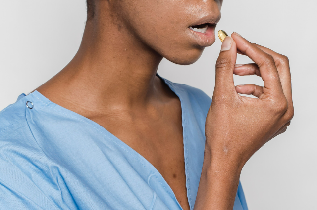
- Patient was advised to take “Sariva Ghana vati‟ (hemidesmus indicus)-250mg (Two tablets three times a day) internally and “Dhara sweda‟(fomentation) was done daily for 20 minutes (except the day of Leech therapy) with lukewarm Teel oil (sesamum indicus at 98.6°F temp) adjuvant to Leech therapy.
OBSERVATIONS
Parameters of observation included Ankle flare, Peripheral Hyperpigmentation, Size of ulcer, Granulation tissues, and relief in Pain. Patient was observed on the above parameters every week for 5 weeks
With Leech Therapy‟ and adjuvant management, the wound completely healed within 30 days i.e. patient was cured from non-healing ulcer
Vrana shodhana and ropana effect: After Leech application expulsion of impure blood takes place, due to which local vitiated doshas (toxins and unwanted metabolites ) are removed. Similarly, it facilitates fresh blood supply and promotes wound healing by the formation of „Healthy Newer Tissues‟. Effect of adjuvent therapy:
- “Teel oil‟ fomentation improves blood circulation, corrects skin discoloration and pacifies venous valvular dysfunction. Thus, it breaks the pathogenesis of „varicosity‟ at the cellular level and helps in wound healing.
- ‘Yashtimadhu Ghrit’ has both „Vedanashamak’ and vran ropan’ properties. Hence, it helps in Healing of wounds and relieves pain too.
- ‘Sira’ and ‘Snayu’ are the bi-product (updhatu) of Rakta and ’ Sariva Ghanvati’ has
‘Raktaprasadniya’ character. Hence, it facilitates the formation of Healthy Newer tissues and also strengthens the blood vessels, thus corrects venous valvular dysfunction
As per Ayurvedic texts, “Sariva’ purifies the Raktadhatu due to its Raktaprasadniya’ character.
Further, once “Rakta’ is purified, its bi-product (updhatu) i.e. „Sira‟ (veins), and its kinematics also get pacified, thus may correct venous valvular dysfunction when used internally along with adjuvant therapy. However, a multi-centric comparative clinical trial along with detailed studies is needed to evaluate the impact of “Leech Therapy‟ on promoting wound healing with respect to varicose ulcer.
Discussions and validation of the above therapy based on Allopathic treatment methodologies.
Non-operative Management The most important aspect of treating patients with non-healing wounds is to address the underlying processes that contribute to the chronicity of the wounds. If the causal factors are not alleviated, wounds will have little chance of healing. In this section, we will discuss the general treatment strategies for each of the common types of chronic wounds (pressure ulcers, diabetic foot ulcers, venous ulcers, and arterial insufficiency ulcers). We will also discuss some basic principles of wound management, including wound debridement, wound dressings, nutritional supports, glycemic control, and adjuvant therapies.
I) General treatment strategy for pressure ulcers
The underlying contributing factors for pressure ulcers must be reduced by providing pressure redistribution and specialized support surfaces. A repositioning schedule to avoid pressure on the wound should be established. Typically, the patients should be repositioned every two hours. Supporting surfaces such as foam mattresses, overlays or air-fluidized beds should be used depending on available resources. The patient’s underlying medical conditions that may contribute to the delay in wound healing should be addressed. Patients should undergo intense physical therapy to help alleviate their immobility status. Systemic infections should be treated and nutritional status optimized. For stage 1 and stage 2 ulcers, treatment should focus on prevention and local wound protection. For stages 3 and 4, treatment usually consists of local ACS/ASE Medical Student Core Curriculum Non-Healing Wounds American College of Surgeons Division of Education Page 18 of 36 Blended Surgical Education and Training for Life® wound debridement followed by meticulous local wound care. While most patients can be managed successfully without surgery, some patients will require delayed closure, skin grafts and/or flaps for coverage. urinary diversion procedures are rarely required and may be associated with higher surgical risks as these patients are already debilitated and malnourished.
Appropriate psychosocial support should also be provided to patients and their families.
II. General treatment strategy for diabetic foot ulcers
Factors contributing to the chronicity of diabetic foot ulcers, such as infection, neuropathy, ischemia, and bony foot deformities should be systematically addressed. Infected wounds should be treated with sharp surgical debridement together with systemic antibiotics. Patients with signs of arterial insufficiency should undergo non-invasive vascular evaluation and/or angiography, and if indicated, revascularization procedures. Mechanical off-loading with total contact cast or cast walkers should be considered to reduce pressure on the ulcers. In patients with foot deformities, referral to a foot and ankle specialist is appropriate as surgical corrections may be required. The recurrence rate for diabetic foot ulcers is extremely high. Therefore, close follow-up and recurrence prevention strategies should be employed. Preventive strategies, such as protective footwear, good foot care, and daily foot inspection have been shown to reduce recurrence in patients with diabetic foot ulcers.
III. General treatment strategy for venous ulcers The primary objective in the treatment of venous ulcers is enhancing venous blood flow. This can be achieved mechanically and pharmacologically. Leg elevation above the heart can improve cutaneous microcirculation, reduce edema, and promote wound healing. Plantar flexion exercise has been shown to improve venous circulation, but its impact on ulcer healing is not known. Nevertheless, this exercise should be recommended to the patients. Static compression therapy with hosiery or bandages is essential in the treatment for patients with venous insufficiency and venous ulcers. Randomized controlled trials have repeatedly demonstrated the benefits of long-term compression therapy in patients with venous insufficiency associated with venous ulcers. These studies demonstrated that venous ulcers heal more quickly with compression therapy than without. Elastic compression therapy (stockings or bandages) is more effective than inelastic compression (Unna boot), based on a meta-analysis. Systemic treatment with aspirin or pentoxifylline (phosphodiesterase inhibitor) has also been shown to improve healing of venous ulcers and should be part of the treatment plan. The selection of wound dressing depends on the level of exudate of the wound and should be individualized. There is no data to support the use of one dressing type over another. Dry, itching and eczematous skin are common in patients with venous disease, and treatment with skin moisturizer should be adequate. If needed, a low to mid-potency topical corticosteroid can be used. Although most patients with venous ulcers can be managed without surgery, in selected patients with large wounds or wounds refractory to medical treatment, a split-thickness skin graft for coverage may be considered. To date, there is no evidence demonstrating the superiority of surgical management over medical management
IV. General treatment strategy for arterial insufficiency ulcers Arterial insufficiency ulcers often begin as minor traumatic wounds that fail to heal as a result of insufficient blood supply to meet the increased demands of the healing tissue. The main strategy in treating arterial insufficiency ulcers is to restore blood flow to the tissue to support wound healing. This can be accomplished by endovascular interventions and/or open vascular reconstructions. Prior to revascularization, an anatomical roadmap of the arterial system should be obtained by computed tomography angiography or digital subtraction angiography. If revascularization is not an option or if revascularization fails, amputation may be the only remaining option. For patients with dry gangrene or with non-infected ulcers, the extremity should be re-vascularized first. Restoration of blood flow is crucial to infection control and must be addressed first prior to any attempt at debridement. In patients with wet gangrene or abscess, the wound should be immediately debrided regardless of any need for revascularization. Once the infection is controlled, a revascularization procedure should follow. Once blood flow has been restored, the focus can then be shifted to local wound care to allow healing, delayed wound closure, and/or coverage. Long-term maintenance of patients with arterial insufficiency ulcers primarily involves risk factor reduction strategies such as smoking cessation and control of diabetes, hypertension, and hyperlipidemia.
Basic principles of wound care management For proper wound healing, the wounds need to be clear of infections, free of necrotic tissue, adequately perfused, and moist. Meticulous local wound care with debridement and proper wound dressings will help maintain a healthy environment that will promote granulation tissue When a wound demonstrates healing potential as evidenced by the presence of granulation tissue and epithelization, it is ready for primary closure or coverage. Wound Debridement: Wound debridement can be accomplished by sharp debridement, wound irrigation, autolytic debridement, enzymatic debridement, and/or maggot debridement. The goal of debridement is to remove infected or devitalized tissue, pathogens, contaminants, and foreign materials, in order to prepare the wound bed for optimal healing and closure. For infected wounds or wounds with large amounts of nonviable tissue, sharp debridement is the most appropriate choice (Figure 8). Sharp excisional debridement reduces the bacterial load and stimulates granulation, contraction, and epithelisation. It is the most rapid and effective way to achieve a clean wound. A disadvantage of sharp debridement is that it causes pain. In cases when significant debridement or pain is expected, it is best to do the debridement in the operating room under anesthesia.
- Infected chronic pressure-injury wound over the left pannus.
- The infected wound has been debrided to remove infected and necrotic materials, leaving a healthy wound base
Wound irrigation can be used to the remove loose necrotic materials from the wound bed and to reduce bacterial load. It should be a routine part of wound management. For most wounds, low-pressure irrigation using a bulb syringe should be adequate. For highly contaminated wounds, high-pressure pulse irrigation should be considered. Sterile saline is the most common solution used for irrigation, but clean tap water can be just as effective. Several studies reported no significant difference in the rate of infection or wound healing rate between clean tap water and sterile saline when used for wound irrigation . It is not necessary to add antiseptics, such as iodine, chlorhexidine, or hydrogen peroxide to the irrigation fluid as they can cause irritation to the tissue and do not add any benefit.
Autolytic debridement is a process in which the body’s own proteolytic enzymes within the wound fluid help break down the devitalized tissue over time. This process facilitates the separation of necrotic tissue from the healthy wound bed to promote wound healing. Autolytic debridement can be augmented by the use of semi-occlusive dressings such as transparent films, hydrogels, and/or hydrocolloids, to keep the wound fluid in constant contact with the wound (see Wound dressings below). A disadvantage of autolytic debridement is that it takes time. Additionally, anaerobic growth may occur with the use of occlusive dressings, requiring frequent wound monitoring for infection. This technique should only be used in non-infected wounds with a minimal amount of devitalized tissue. Enzymatic debridement involves the application of commercially available enzymatic agents such as collagenase or papain to the wound. Collagenase is an enzyme isolated from the bacterium Clostridium histolyticum. It possesses the ability to selectively digest collagen in necrotic tissue but not in healthy tissue. Papain is a proteolytic enzyme that is found naturally in the papaya fruit. Its proteolytic function must be activated by urea which is also included in the commercially available papain formulation. Another papain ointment formulation contains papain, urea and chlorophyllin. Similar to collagenase, papain selectively digests proteins in nonviable tissue and spares the healthy granulation tissue. Papain should not be used in the presence of hydrogen peroxide, which may inactivate the enzyme. It is the general consensus that enzymatic debridement and autolytic debridement are slow and are only effective in wounds with minimal necrosis. They can be used as an adjunct to surgical debridement. Medical maggot therapy has been used in the treatment of chronic wounds. Maggots secrete proteolytic enzymes that break down necrotic tissues which are then ingested by the larvae leaving behind healthy tissue. There is data to suggest that maggot therapy in addition of conventional wound care results in more complete debridement compared to conventional wound care alone. However, this has not translated into reduction in time to complete wound healing in randomized controlled trials. Pain can be associated with maggot therapy, but the main disadvantage of maggot therapy is the negative perception about its use by patients and staff. Wound dressings: Local wound care with the help of wound dressings is an important element in the preparation of the wound bed for healing, wound closure, skin graft, and/or flap closure. The choice of wound dressings can have an impact on the speed of wound healing and potentially the cosmetic appearance of a healed scar. There are several different types of wound dressings to choose from; each has its unique characteristics. Unfortunately, there is no single type of dress that can be used in all situations. The type of wound dressings used has to be individualized, based on the characteristics of the wound and the stage of healing. Moisture level on the wound bed is very important for wound healing. The wound fluid is rich in platelet-derived growth factors, fibroblast growth factors, and metalloproteases that are important in wound healing. The moist environment is also thought to promote the migration of epithelial cells required for reepithelization. Studies in both animal models and human models have shown that moist wounds heal faster than dry wounds. The primary purpose of wound dressings is to control the moisture level on and around the wound. The wound should be moist enough to promote wound healing, but excess exudate must be controlled to prevent maceration of healthy tissue. Thus, this moisture/exudate balance is the basis for the individualized choice of wound dressings. For example, high-absorbent dressing, such as alginates and foams, should be used in high-exuding wounds, while hydrogels, with the ability to donate water, should be used in dry/low-exuding wounds. The stage of the wound is also a consideration for dressing selection. During the debridement stage, a dressing that can perform mechanical debridement or promote autolytic debridement is preferred. During the granulation stage, low adherent moisture-retaining dressing will help protect the delicate granulation tissue, while maintaining a moist environment for epithelial migration. Common types of wound dressing materials are discussed below.
- Gauze dressing is the most common dressing used in open surgical wounds. The gauze is moistened with saline prior to placing it into the wound. Typically, more absorbent gauze or pads are placed over the moist gauze and secured by tape or bandage. As the moistened gauze dries, it adheres to the surface of the wound, so when the dressing is removed during dressing change, some of the devitalized tissues come with it. This method of wound care is commonly known as wet-to-dry dressing. This is typically used on infected wounds, freshly debrided wounds, or wounds that still have a lot of devitalized tissues. The dressing is usually changed twice daily. If the wound develops pseudomonas colonization as evidenced by the presence of blue-green necrotic debris or if bacterial colonization increases as evidenced by the presence of odor, the dressing solution can be changed to Dakin’s solution (0.25% to 0.5% sodium hypochlorite or bleach). Advantages of gauze dressings are that they are inexpensive, readily available, and they can be used virtually on any type of wound. Disadvantages of gauze dressings are that they can debride developing granulation tissue and that they do not foster a moist wound healing environment. For these reasons, wet-to-dry gauze dressings should be discontinued when necrotic tissue is gone and granulation tissue is present.
- Impregnated gauze dressing is another commonly used dressing. It is fine mesh gauze impregnated with petroleum, paraffin wax or antibiotic ointment. The primary mesh dressing is applied directly to the wound, which is then covered with another layer of absorbent pads. These dressings are non-adhering so that they can be easily removed during dressing changes with little pain or trauma to the regenerating tissue. On the other hand, these dressings do not maintain a moisture-rich environment or provide good exudate control. Fluid can easily go through the mesh gauze and can collect in the overlying pads, causing desiccation of the wound bed and maceration of the surrounding skin.
- Transparent films are sheets of self-adhesive occlusive dressings that are permeable to water vapor and oxygen, but impermeable to larger molecules such as proteins and bacteria. This property allows oxygen to enter into the wound, and at the same time, allows moisture vapor to escape. These films trap wound fluid within the dressing and maintain a moist wound healing environment, while simultaneously preventing bacteria invasion. These films can stay in place for up to one week. Because they are transparent, they do not have to be removed for wound inspection. These dressings are typically used on partial-thickness wounds such as donor sites and superficial wounds such as minor burns. These films do not have any absorptive capacity and therefore should not be used on moderately or heavily exudative wounds. The leakage of wound fluid to the surrounding skin can cause maceration. Sometimes the adhesive sticks to the wound bed and removal can be painful.
- Foam dressings are composed of a hydrophilic foam inner layer that conforms to the wound bed and a hydrophobic gas-permeable outer layer to prevent leakage and bacteria contamination. Some foam dressings are self-adhesive, while others require a secondary adhesive dressing. They are commonly used on pressure ulcers, minor burns, skin grafts, diabetic ulcers, donor sites, and venous ulcers. The foam layer is soft and does not adhere to the wound bed, making it more comfortable for the patients. They are highly absorbent and should be used in moderate to highly exuding wounds. They should not be used in dry or low-exuding wounds as desiccation can occur.
- Alginate dressings are made from calcium alginate, a polysaccharide salt derived from algae. When it comes in contact with sodium-rich wound fluid, the exchange of calcium for sodium irons results in the formation of a strong hydrophilic, highly absorbent gel that conforms to the wounds. Alginate dressings come in various forms, including ribbons, beads, and pads. Their highly absorbent characteristic allows effective control of exudates while maintaining a moist wound bed. Because the dressing can conform to the shape of the wounds, they can be used for packing deeper wounds or wounds with tunneling. Alginates are nonadherent and can easily be washed away with saline, causing minimal pain to the patients. They should not be used in dry or low-exuding wounds, or wounds with tendon or bone exposed, as they can cause desiccation. Alginate dressings usually require secondary dressings.
- Hydrocolloid dressings are made up of colloid particles such as gelatin, pectin, and carboxymethylcellulose that are carried on self-adhesive polyurethane films, forming flexible wafers. When the colloid material comes in contact with exudate, it can swell into a gel-like mass. The colloid material is moderately absorbent, so it can be used in moderate exuding wounds, but not in highly exuding wounds. The colloid is impermeable to water, traps wound fluid and promotes moist wound healing. It is also impermeable to bacteria and potentially can be used in areas of high risk for stool contamination (near ostomy site). These dressings are commonly used on burns, pressure ulcers, and venous ulcers. On the other hand, the colloid can also trap bacteria inside the wound, so it should not be used on infected wounds. The dressings are usually changed daily and they can leave a residue on the wound bed.
- Hydrogels consist of a matrix of synthetic polymers that is capable of holding a large amount of water. In the gel-base form, hydrogels are composed of more than 90%. Hydrogel dressings come in either free-flowing gels or hydrogels impregnated into gauze pads, sponge ropes, strips or fine mesh sheets. Hydrogel matrices can absorb or donate water depending on the hydration state of the surrounding tissue. Hydrogels are most useful in dry or dehydrated wounds. The cooling sensation provided by the hydrogels can offer some pain relief in some patients. Hydrogels have been found to selectively permit gram-negative bacteria to proliferate, but no increased risk of infection has been observed. They should be avoided in infected wounds or wounds with high risk of bacterial infection.
- Topical antimicrobial agents: There are some topical antimicrobial agents that have been used for chronic wounds. These include iodine-based agents (cadexomer iodine), silver-based agents, and medical-grade honey. Both iodine and silver are toxic to bacteria and, theoretically, can reduce bacteria load within the wound and promote wound healing. Interestingly, iodine-induced hyperthyroidism has been reported in patients treated with cadexomer iodine. Honey is toxic to bacteria due to its high osmolarity and high hydrogen peroxide concentration.
Operative Management
- Delayed wound closure It is important to stress that the underlying etiology of chronic wounds should be addressed prior to performing the definitive closure. The wounds also have to be clear of infections, free of necrotic tissue, and adequately perfused before any attempt at delayed closure. The wounds have to be small enough to allow closure with minimal tension. The wound edges are sharply debrided to allow better tissue approximation. Vertical mattresses with monofilament no absorbable sutures are usually used to bring the deep and superficial edges together.
- Split-thickness skin grafts Split-thickness skin grafts (STSGs) are autologous dermal grafts that consist of the epidermis and a part of the dermis. Split-thickness skin grafts are harvested by a dermatome at a thickness that can be pre-adjusted on the instrument by the surgeon. The thickness of the grafts can range from 0.008 to 0.012 inches. Split-thickness skin grafts can be harvested pretty much anywhere, but the most ideal places for harvest are large surfaces of thick skin such as the thighs and torso. Once the graft is harvested, the wound at the donor site is left to heal by re-epithelisation, taking advantage of the remaining hair follicles and sebaceous glands to serve as a source of epidermal progenitor cells.
a. Split-thickness skin graft of the left upper extremity. The graft was meshed with a ratio of 1:2. The photograph was taken one day after surgery.
b. A healed split-thickness skin graft of the left lower extremity 6 months after surgery.
The graft is usually meshed to allow expansion over a larger area. The meshing of the graft involves using a meshing device to make rows of short interrupted cuts of a few millimeters long. This allows the mesh to stretch over an area that is larger than the area of the original harvested
Conclusion with a take-home message
From the above discussions and research carried out, With “Leech Therapy‟ and adjuvant Ayurvedic treatment, the non-healing varicose ulcer completely healed within 30 days. Validation has been carried out based on extensive experimentation and based on allopathic treatment methods widely used in this field. On the basis of this case study, we can roughly conclude that Ayurveda can give a ray of hope in the treatment of varicose veins and ulcers. None of the complications like severe bleeding, wound infection, or hypersensitivity were observed during the therapy. Leech therapy‟ proves to be effective, time-saving, affordable and acceptable treatment. Though treating non-healing “Varicose ulcer‟ is a difficult task, we have managed to treat it with „Leech Therapy‟ along with conventional (Ayurvedic) methods of wound care. A multi-centric comparative clinical trial along with valvular studies is needed to establish this unique treatment protocol.
. . .
Bibliography
REFERENCES:
- http://en.wikipedia.org/wiki/Venous_ulcer date 25/04/2013
- Bush, R. New technique to heal venous ulcers: Terminal interruption of the reflux “Teel oil’ Dhara swed‟ in Varicose ulcer
- Prognosis on Day 30 Dwivedi Amarprakash: Management of Non-Healing Varicose Ulcer In Ayurveda: Perspectives in Vascular Surgery and Endovascular Therapy.
- Dr. Anantram Sharma, „Sushrut vimarshini‟ commentary on Sushrut Samhita, Chikitsa sthan - Chapter 4 (Vat Vyadhi chikitsa- Shlok 7) Volume 2, Published by Chaukhambha Prakakashan2009, Page 205
- Illustrated Sushrut Samhita, Translated by K. R. Srikant Murthy, Second edition: 2004
- Medicinal Leech Therapy, Author: Andreas Mechallsen, Manfred Roth, Gustav Dobos. Publication-Theme, New York, USA, 2007.
- Weinfeld AB et al, Clinical and Scientific consideration in Leech therapy for the management of acute venous congestion,
- Amarprakash P. Dwivedi, Case Study of Leech Application in Diabetic foot ulcer, IJRAP (International Journal of Research in Ayurveda and Pharmacy), Volume 3, Issue 5, Sep-Oct 2012